CEA-Paris-Saclay is currently working on an MRI project to revolutionize the medical field. The researchers recently obtained very promising first images. However, the use of this MRI on a pumpkin raises questions. What is it really?
This is about the most powerful MRI in the world intended for imaging in humans, as explained by Stanislas Dehaene in a press release from CEA-Paris-Saclay of October 7, 2021. He directs the French neuroimaging platform Neurospin and refers to the Iseult project coming all just to prove yourself. According to him, this marvel of engineering draws its strength in its magnet .
It must be said that its characteristics are very surprising. The device weighs 132 tons and measures 5 m in length, 5 m in external diameter for an internal diameter of 90 cm. It has a nominal magnetic field of 11.7 Tesla, which is an all-time high. Indeed, the usual hospital MRIs count only 1.5 to 3 Tesla. In addition, its field is powered by an electric current of 1,500 amps . This power supply is obtained via conductor coils permanently cooled by helium in a superfluid state at -271.35°C.
The magnet in question is nothing more than a very high resolution imager and will therefore be used to scan images. The CEA recently unveiled the first results of the MRI, whose incredible resolution is 400 microns in all three dimensions (see below).
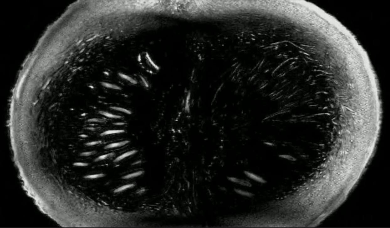
This very serious research also has an unusual dimension. Indeed, project researchers used a pumpkin to get their first images. And yet, this choice was not made by chance. It turns out that the consistency of this vegetable as well as its size have common points with our brain , in addition to having multiple textures. In addition, the pumpkin contains about 90% water, a value quite close to our brain (80%).
Thus, having used a pumpkin as a guinea pig made it possible to deliver a real proof of concept, demonstrating the feasibility of this innovation . The researchers believe that the images are promising and now hope to optimize their device to obtain a resolution of 100 or 200 microns . Although this figure is lower than the 400 microns of the first images, it will achieve a higher effective resolution, due to better precision.
This is a revolution for the medical field. Specifically, this innovation should support basic research , work in the cognitive sciences as well as knowledge of cerebral pathologies such as Alzheimer's or Parkinson's. This will also speed up medical diagnoses. The next step for the scientists will be to test their equipment on humans, obviously after the green light from the health authorities .