Various techniques can be used for physical examination. The most important techniques for looking at your body from the inside at a glance:ultrasound, X-ray CT, PET and MRI.
Echo Ultrasounds are mainly made for abdominal complaints (and during pregnancies). This is done with sound waves of a high frequency. The waves are reflected by the body tissues and converted into a video image.
X-ray To get an image of, for example, bone fractures, X-rays are made. The X-rays pass through the body and give a kind of shadow image of the organs and the skeleton. An X-ray can also provide more clarity when heart failure, pneumonia or a lung tumor are suspected.
CT (Computer Tomography)
A CT scan also uses X-rays, but cross-sections are now made that are translated by the computer into 3D images. In this way all organs, but also abnormalities in bones or tissues can be viewed.
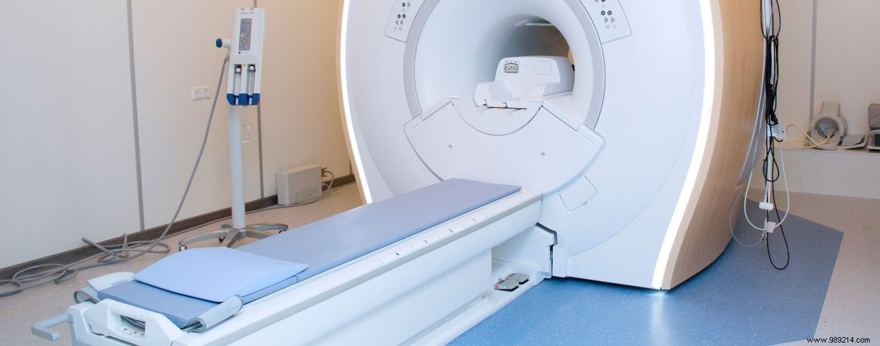
MRI (Magnetic Resonance Imaging)
Does not work on the basis of radiation, but with radio waves. The patient lies in a tube containing a very strong magnet that makes the body magnetic. Because the chemical composition of tissues varies, their magnetic properties are also different. These differences allow images to be created. The images can be viewed with the computer as 'slices' of the body. MRI is a useful tool, especially in brain research.
PET (Positron Emission Tomography)
The body is injected with a radioactive substance. The substance spreads throughout the body and emits radiation. By measuring the radiation with detectors, images can be made. Recent research has shown that a PET scan can detect infections and foci of inflammation better than other techniques.